|
|||||||||||||||||||||
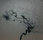
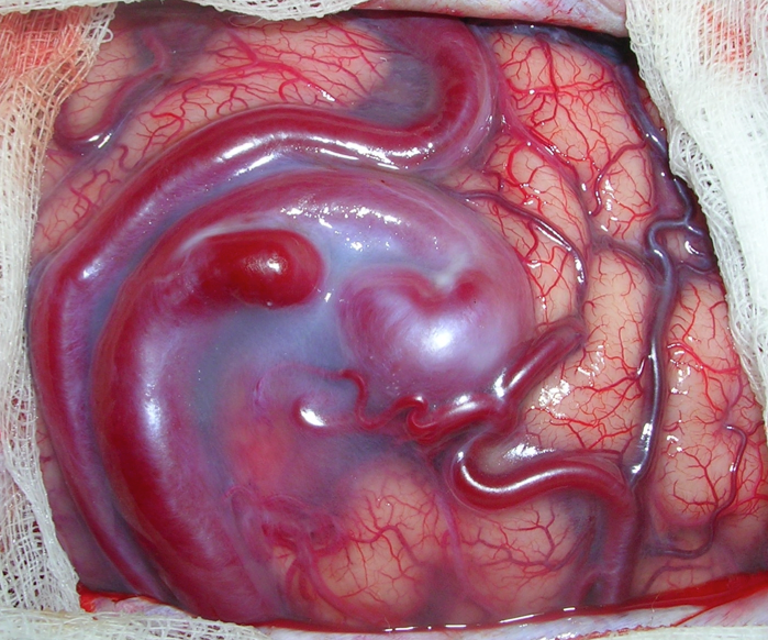
A 23-year-old lady, previously well, complained of sudden, severe headache, dizziness, and vomiting. She was rushed to the nearest emergency room for treatment. On examination, the patient was drowsy but able to follow commands. CT scan of the brain showed a blood clot in the right occipital area (back part of the brain). Because the patient was young and did not have other medical problems, an angiogram was requested to investigate the source of the bleed. The angiogram showed an arteriovenous malformation (AVM) in the right occipital area. An AVM is an abnormal tangle of blood vessels. They are believed to be congenital, which means that a patient is born with this condition. AVM’s may present in different ways, such as bleeding or seizures, but some may not cause symptoms at all. Management depends on several factors such as the patient’s age, size of the AVM, location of the AVM, and others. This patient underwent surgery for excision of the AVM. Upon opening, large, angry-looking vessels were seen on the surface of the brain (please see picture). These were traced to the body of the AVM, and the entire AVM was removed to prevent it from causing another bleed. The patient recovered well from the surgery. Prior to the surgery, she already complained that she could see only half out of each eye, as if there was a curtain blocking half of her visual field. This was due to the location of the bleed and the AVM in the occipital lobe, the part of the brain that controls visual processing. The visual problem persisted after surgery, but it is expected to improve over time. |
|
|
|
|
|||||||||||||||||